

A new type of brain scan is showing promise as a powerful tool for assisting surgeons in maximising removal of cancerous tissues.
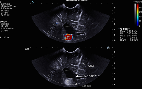
Known as shear wave elastography, the technology is a type of ultrasound that measures the stiffness and elasticity of tissue. Vibrations – the ‘shear waves’ – are passed through the subject, detecting the relative stiffness of different brain matter. On average, brain tumours tend to be stiffer than normal brain tissue, and the scan works by mapping suspicious areas of particular stiffness.
VHEE beam makes precision strike on tumours
AI algorithm detects and identifies brain lesions
Unlike MRI scans – the gold-standard for cancer detection – sheer wave scans can be carried out in theatre, with the information passed immediately to the surgeon to enable the removal of residual cancerous tissue without additional surgeries. In a study published in Frontiers in Oncology, it was found the technique outperformed surgeons at detecting leftover cancerous tissue by sight alone, and could prove to be a vital technology in improving outcomes for brain tumour patients.
“Ensuring all of a brain tumour is removed without damaging healthy tissue is a major challenge in brain surgery,” said study lead Professor Jeffrey Bamber, Professor in Physics Applied to Medicine at The Institute of Cancer Research, London. “Using this new type of scan, surgeons could greatly increase confidence that no cancerous tissue is going to be left behind after surgery.”
The study was led by The Institute of Cancer Research and the National Hospital for Neurology and Neurosurgery. In a sample of 26 patients, it compared the efficacy of three different techniques: shear wave scans, a standard 2D ultrasound, and a surgeon’s opinion. It found that shear wave elastography was more sensitive in detecting residual tumour tissue than a standard ultrasound or the surgeon alone.
Shear wave scans detected tumour tissue with 94 per cent sensitivity, compared with 73 per cent for standard ultrasound and 36 per cent for the surgeon. This means that when there was residual tumour material, shear wave scans were 2.5 times better than the surgeon at detecting it.
However, shear wave scans detected tumour tissue with only 77 per cent specificity – better than the 63 per cent for standard ultrasound but less effective than the 100 per cent for surgeons. These numbers suggest the new technique could yield more false positives than surgeons acting alone, and the researchers believe the technology will be best used to augment a surgeon’s opinion.
“Imaging plays a crucial role in many aspects of cancer treatment, in providing valuable information about tumours and ensuring doctors don’t have to make decisions blind,” said Professor Kevin Harrington, head of the Division of Radiotherapy and Imaging at The Institute of Cancer Research, London.
“This new study has shown for the first time that a particular type of ultrasound scan could provide real-time guidance to brain surgeons during operations as they choose which tissue to remove. It’s an exciting area of research which has the potential to improve outcomes for patients by ensuring surgeons take out the entire tumour while minimising damage to the healthy brain.”
Shear wave scans were also shown to be as good as post-surgery MRIs at detecting tumour tissue that had been left behind, making them a cheaper, faster and more feasible alternative. According to the research, larger studies will now be required to confirm the results of the work.
The post New brain scan tech could improve tumour removal appeared first on The Engineer.